Clinical Example: Proximal Interphalangeal Joint Flexion Contractures after Dislocations and Reflex Sympathetic Dystrophy
| Reflex sympathetic dystrophy is most often dealt with as a painful problem complicating recovery. It may resolve or become chronic with or without treatment. Active RSD has components of vasomotor instability, pain, and avoidance. Structural changes, such as arthrofibrosis and soft tissue atrophy may develop and persist. |
| Click on each image for a larger picture |
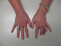
| Four years ago, this
patient sustained closed middle finger PIP dislocation a ring
finger intraarticular middle phalanx base fracture. She developed
painful reflex sympathetic dystrophy. PIP and DIP joint stiffness
developed during the painful phase and remained after resolution
of all other RSD related issues. |

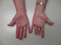
| Range of motion was
limited: middle PIP fixed at 90 degrees; ring PIP fixed at 80. The
middle DIP is stiff at 0 degrees; the ring DIP ranges only from 0 to 20
degrees hyperextension. |

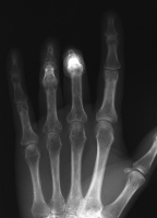
| Standard AP radiographs
fail to demonstrate PIP joint anatomy because of contractures. |

| Modified AP views taken
with the middle phalanges flat on the radiograph plate show
preservation of joint anatomy. |

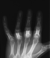
| Oblique view. |

| Lateral view shows PIP
joint space narrowing, more pronounced in the ring finger. They also
show periarticular osteoporosis of the middle and ring fingers compared
to the index and small - impressive in that this is four years
post injury and the patient uses the fingers normally within the
constraints imposed by contractures. |

| Search
for... RSD RSD stiffness |
Case Examples Index Page | e-Hand home |